

Generated using ChatGPT (GPT-4)
Forgetting an appointment or misplacing your keys can be frustrating. Forgetting how to get home from a familiar grocery store, how to prepare a favorite meal, or why a loved one’s face seems unfamiliar is something else entirely. For people living with Alzheimer’s disease, memory loss can slowly turn ordinary routines into daily obstacles. This disease erodes not only memory, but also corrupts independence, identity, and the small habits that make life feel recognizable.
A Disease That Disproportionately Affects Women
In Canada, an estimated 771,939 people were living with dementia in 2025. Women carry a disproportionate share of that burden: in 2020, an estimated 61.8% of Canadians living with dementia were female. Women face an estimated 1 in 5 lifetime risk of Alzheimer’s disease, compared with 1 in 10 for men, yet female biology has often been treated as a complication in brain research: something that makes experiments harder to control rather than something worth understanding. A systematic review and meta-analysis of 56 Alzheimer’s randomized clinical trials involving 39 575 participants found that women made up 59% of participants despite a much higher proportion of women with Alzheimer’s in the general population. Furthermore, only 12.5% of studies reported results separately by sex. This limited reporting makes it difficult to determine whether disease progression, treatment effectiveness, or adverse effects differ in men and women, illustrating how overlooking sex differences can leave clinically important aspects of neurological disease undetected.
Much of this perceived complexity comes from fluctuations in ovarian hormones such as estrogen and progesterone across the menstrual cycle, pregnancy, and menopause. These hormones decline with age, dropping significantly during menopause. They can influence how neurons get excited, how the brain maintains and strengthens connections between neurons, metabolism, inflammation, and more crucial neural processes. Researchers have historically treated female variation as experimental noise that was easier to avoid rather than study. As a result, male animals and male physiology quietly became the default, even though female hormonal changes may themselves be important to understanding sex differences in Alzheimer’s disease.
What Changes When Hormones Change?
What happens when researchers stop treating the female brain as a complication, and start asking whether hormonal life stages change the biology of Alzheimer’s disease? A 2024 study led by researchers at Western University and collaborating institutions helps show why that question matters. Led by graduate student Liliana German-Castelan under the supervision of neuroscientist Vania Prado at Western University’s Schulich School of Medicine & Dentistry, the research team also included collaborators from the RIKEN Center for Brain Science in Japan. For the human portion of the study, the researchers used data from the Australian Imaging, Biomarkers and Lifestyle study of ageing. The paper was first published online in October 2023 and appeared in the 2024 volume of Alzheimer’s & Dementia.
The team examined two major players in Alzheimer’s disease. The first was amyloid beta, a protein that can build up in the brain and form plaques, one of Alzheimer’s most recognizable features. The second was acetylcholine, a chemical messenger that helps brain cells communicate and supports attention, learning, and memory. Brain cells do not touch each other directly. Instead, they pass chemical messages across tiny spaces between them, a bit like passing a note across a narrow gap. Acetylcholine is one of the chemicals that helps deliver those messages. Because it supports attention, learning, and memory, scientists have long been interested in what happens when this communication system begins to weaken in Alzheimer’s disease.
The researchers asked whether weakening this acetylcholine system would affect amyloid buildup differently in male and female brains. They also asked whether estradiol, the main form of estrogen before menopause, might help explain any differences.
From Mice to Human Brains
To investigate this question, the researchers combined experiments in mice with an analysis of human brain scans. They used mice with Alzheimer’s-associated genetic mutations and, using gene therapies, altered the amount of acetylcholine their brain cells could release. They did this by changing levels of the vesicular acetylcholine transporter, a protein that helps package acetylcholine for release between brain cells. Some female mice also had their ovaries removed so the researchers could examine how loss of ovarian hormones (mimicking menopause) affected these relationships. The researchers then measured amyloid beta in male and female brains.
For the human portion, the team analyzed repeated structural MRI scans, which show changes in brain tissue, and amyloid PET scans, which reveal amyloid buildup, from older adults participating in the Australian Imaging, Biomarkers and Lifestyle study of ageing. MRI allowed them to track tissue loss in the basal forebrain, a region containing many acetylcholine-producing neurons, while PET imaging showed how much amyloid had accumulated in the brain. By following these measures over time, they could test whether deterioration of the cholinergic system was associated with increasing amyloid in men and women.
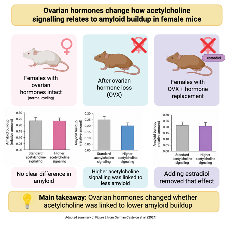
Figure 1: Adapted summary of Figure 3 from German-Castelon et al. (2024)
When Ovarian Hormones Disappear, the Pattern Changes
The central finding was that ovarian hormones changed the relationship between acetylcholine signalling and amyloid pathology. Female mice with normal ovarian hormone levels did not show a clear relationship between reduced acetylcholine activity and greater amyloid buildup. After their ovaries were removed, that pattern changed: lower acetylcholine activity became associated with greater amyloid accumulation. Interestingly, the female mice in a menopause-like state began to resemble the male mice, which also showed greater amyloid buildup when acetylcholine activity was reduced. The human data showed a similar pattern, with men and post-menopausal women both showing an association between poorer health of the brain’s acetylcholine system and greater amyloid accumulation over time.
In other words, the relationship between acetylcholine and Alzheimer’s related pathology was not constant across female hormonal states. Loss of ovarian hormones shifted the female pattern towards one that more closely resembled males.
Why the Male Default Can Miss Part of the Story
For decades, many Alzheimer studies relied heavily on male mice. This research shows why this can be a major blind spot. A treatment that works in a male brain may not work the same way in a female brain, especially before menopause. That distinction is important: female biology cannot be treated as a fixed category or as a slightly more complicated version of the male default.
The goal is not to force every study to yield results that show a dramatic difference between men and women. Rather, we should question how the results may differ across the various hormonal stages a woman experiences in her lifetime. We don’t rule out that some findings may be similar, but researchers need to know when they are similar, when they are different, and whether those patterns change across the lifespan.
That is why including women in Alzheimer’s research is not just about comparing “male brains” and “female brains.” It is about recognizing that female biology is dynamic. Puberty, menstrual cycles, pregnancy, perimenopause, menopause, and aging can all alter hormones, metabolism, immune activity, blood flow, and communication between brain cells. Any of these changes could influence when Alzheimer’s-related changes begin, how quickly they spread, how symptoms appear, or which treatments work best.
Original Article: German-Castelan, L., Shanks, H. R. C., Gros, R., Saito, T., Saido, T. C., Saksida, L. M., Bussey, T. J., Prado, M. A. M., Schmitz, T. W., Prado, V. F., & Australian Imaging Biomarkers and Lifestyle flagship study of ageing (2024). Sex-dependent cholinergic effects on amyloid pathology: A translational study. Alzheimer's & dementia: the journal of the Alzheimer's Association, 20(2), 995–1012. https://doi.org/10.1002/alz.13481