Rethinking the “Little Brain”: A New Brain Map Reveals the Surprising Complexity of the Cerebellum
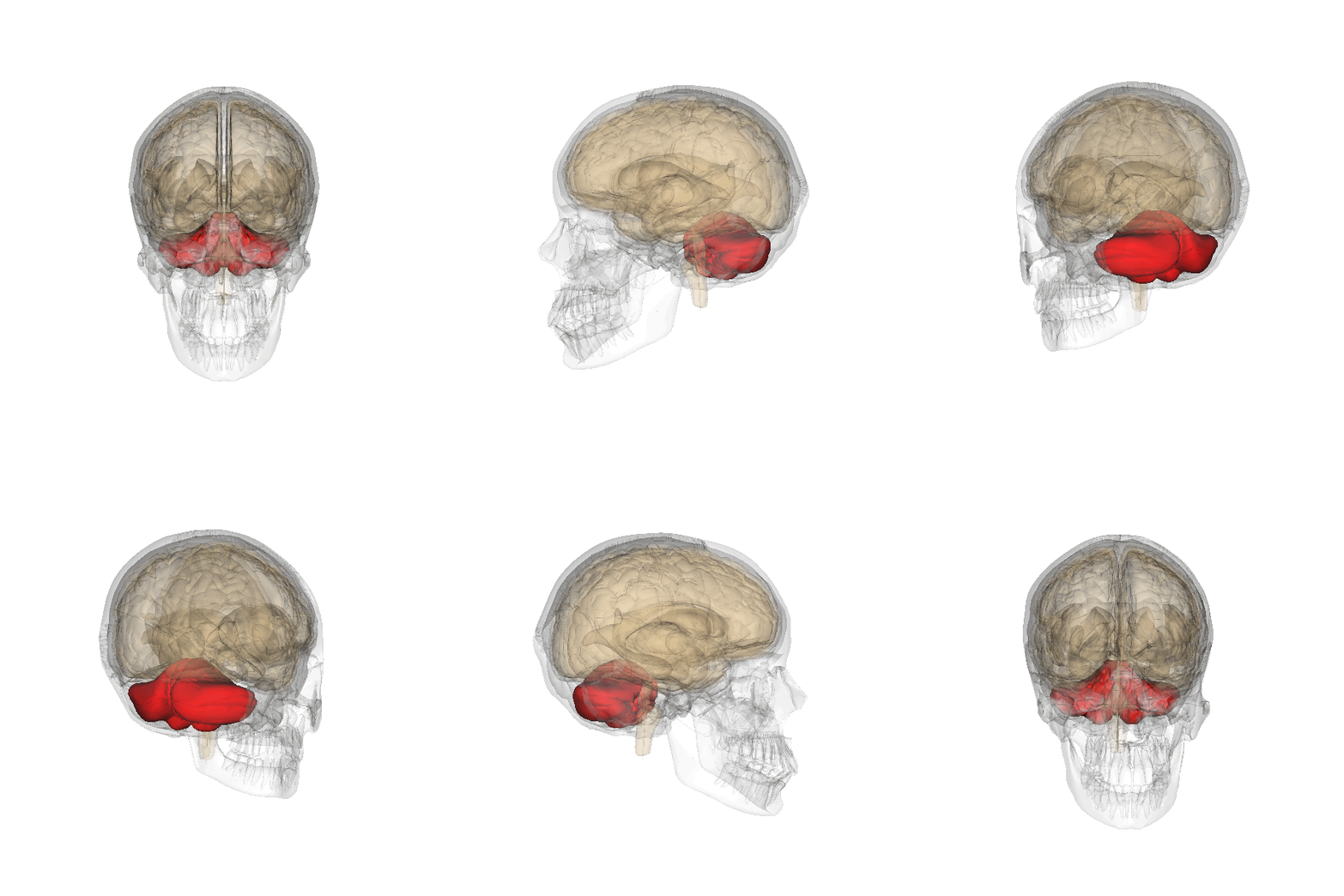
Figure 1: Human cerebellum. Adapted from Life Science Databases (LSDB) Anatomography (BodyParts3D). Licensed under CC BY-SA 2.1 Japan.
Our brain surprises us every day. Despite decades of scientific research, there is still so much we do not fully understand about this remarkably complex organ. Every year, scientists uncover new insights about how the brain works, revealing layers of organization that were previously invisible to us.
One brain region that has undergone a major shift in scientific understanding is the cerebellum. Often referred to as the “little brain,” the cerebellum sits at the back of the brain beneath the cerebral cortex. Although the cerebellum is much smaller than the cerebral cortex, scientists estimate that it contains over 100 billion neurons, which is more than half of all the neurons in the human brain. This is a remarkable feature for a structure once thought to mainly support movement and balance.
Newer evidence has begun challenging this idea!
For many years, scientists understood the cerebellum mainly through observations of patients with damage to the region, who often showed problems with movement, balance, and coordination. Later studies revealed that cerebellar damage could also affect cognition (including thinking and problem-solving), language, and emotion, suggesting that this region might play a far broader role than previously believed. As scientists began uncovering hints that the cerebellum might be involved in more than movement alone, researchers started using brain imaging techniques to map its functional organization. These studies revealed that the cerebellum contains many distinct functional regions, but no single map has been able to fully capture its complexity.
So how do scientists even begin mapping something this complex?
Researchers can use brain imaging tools such as functional MRI, which measures changes in blood flow to reveal which brain regions are active while participants perform specific tasks. Participants might solve problems, remember information, move their fingers, answer questions, or respond to images while researchers monitor which parts of the brain become active. By comparing brain activity across different tasks, scientists can begin building functional “maps” showing how different brain regions contribute to different behaviors and mental processes. Over the years, approaches like these have helped scientists create many of the brain maps and functional atlases used in neuroscience today.
Advances in technology are now allowing researchers to study the brain in much more powerful ways. Traditionally, most brain atlases were built using a single experiment or one specific set of tasks, meaning that each study could only capture part of the brain’s functional organization.
In a recent study, Dr. Caroline Nettekoven and her colleagues took a very different approach to mapping the cerebellum. Instead of relying on one dataset alone, the team combined years of brain imaging data collected across eight large datasets. Seven involved participants performing a wide range of tasks, while the eighth examined how different brain regions communicate while participants were simply resting. Together, these datasets captured brain activity from over a hundred participants performing hundreds of different task conditions designed to probe movement, memory, attention, language, social thinking, and problem-solving. Much like using many flashlights to illuminate different parts of a dark city at night, combining these datasets allowed the researchers to build a far more complete picture of the cerebellum’s organization than any single study could provide.
But combining these datasets was not as simple as stacking them together. Each study was designed differently, with different tasks, different participants, and data collected across different scanning sessions. To overcome this challenge, the researchers used advanced computational methods to identify functional patterns that consistently appeared across all datasets while still accounting for the fact that every individual brain is slightly unique.
The result was one of the most detailed functional atlases of the cerebellum ever created. The atlas was organized hierarchically, meaning scientists could study the cerebellum at different levels of detail, from broad functional systems down to smaller specialized subregions nested within them. Much like Google Maps allows users to zoom from countries to cities to neighbourhoods, this new atlas lets researchers zoom in and out of the cerebellum’s organization, revealing both large-scale functional networks and fine-grained specialized regions hidden within the “little brain.”
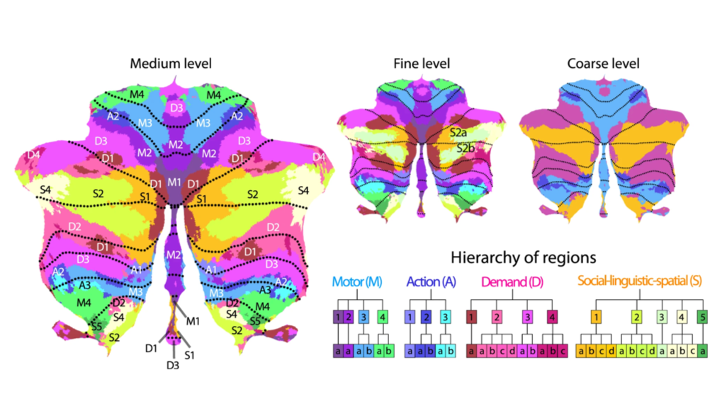
Figure 1. A new functional map of the human cerebellum at different levels of detail. The researchers organized the cerebellum into multiple levels, allowing scientists to “zoom in and out” of its functional organization much like Google Maps. The coarse-level map (right) shows broad functional systems, while the medium- and fine-level maps (left and center) reveal increasingly specialized subregions involved in movement, action, attention, language, social thinking, and other cognitive functions. Regions with similar colors tend to behave more similarly during different tasks. The diagram at the bottom illustrates how these functional regions are related to one another in a hierarchical organization, with larger systems branching into smaller specialized subregions.
The researchers discovered that the cerebellum is organized into distinct functional regions involved in different types of behaviour and thinking. Some regions were strongly linked to movement and body control, while others were associated with attention, working memory, language, social thinking, imagination, and action observation.
Interestingly, each dataset used in the study revealed different aspects of cerebellar organization. Datasets that included detailed movement tasks better revealed motor regions, while datasets involving memory, language, and executive tasks more clearly highlighted regions linked to attention, social thinking, and other cognitive functions. By combining all of these datasets together, the researchers were able to build a more complete and reliable map of the cerebellum than previous atlases.
Perhaps most importantly, the study showed that while humans share a common cerebellar organization, every person’s cerebellum is also slightly unique in its functional organization. This opens the door for more personalized brain mapping in the future and may help scientists better study neurological disorders, brain connectivity, individualized treatments, and how cognitive functions differ across people at a much more precise level.
The cerebellum is small. But this research suggests it may be one of the most intricately organized regions in the human brain. As scientists continue developing new ways to map the brain, an even more complex and fascinating picture is beginning to emerge.
Original Article: Nettekoven, C., Zhi, D., Shahshahani, L., Pinho, A. L., Saadon-Grosman, N., Buckner, R. L., & Diedrichsen, J. (2024). A hierarchical atlas of the human cerebellum for functional precision mapping. Nature Communications, 15(1), 8376. https://doi.org/10.1038/s41467-024-52371-w