
What does the size of my child’s brain have to do with how they develop socially and emotionally?

Microsoft Stock Images
How does the size of different brain areas relate to brain function? An intuitive understanding might suggest that bigger brains in general, or bigger areas, are better at functioning. However, recent research suggests that this may not always be the case, especially when you start looking at specific parts of the brain. For example, a study published in 2025 focused on this question within a part of the brain called the amygdala. Among many functions, the amygdala is broadly involved in regulating our involuntary bodily functions (e.g., reflexes), regulating the autonomic nervous system functions, and the secretion of hormones, processing and assessing the meaning and importance of information from our senses, as well as processing social and emotional information (source). The article discussed here suggested that increased size of the amygdala was associated with worse functional outcomes.
In their study, Mueller and colleagues (2025) narrowed down their investigation by asking how the size of the amygdalae is associated with social and emotional development in infants and children. Understanding how the amygdala changes in a sample of healthy or typically developing children in early childhood provides a reference point or benchmark for understanding what atypical amygdala development looks like. This included participants from several cohorts of healthy fetuses (from 28 weeks pregnant onward), and infants and children aged approximately 6 months to 4 years old. The data used in the study were sourced from several locations, including but not limited to Ontario, Alberta, California, Pittsburgh, Michigan, and England. Sampling across several sites increases the research generalizability of the study, meaning that the findings could apply to a broader population of individuals. The study made use of both longitudinal data (participants were measured at multiple time points) and cross-sectional data (participants were measured at one time point only). See Box 1 for details about longitudinal and cross-sectional approaches.
Box 1. Challenges in Longitudinal Research
A feature of this study is that it included both longitudinal data (having the same individuals do the study multiple times) and cross-sectional data (each individual participates in the study at one time point only). What makes a study longitudinal is that each individual partakes in several sessions of the experiment over an extended period. Some examples of longitudinal studies are completing a series of surveys once a month for a year, doing a memory test 3 times over the course of several months, or having your brain scanned multiple times over the course of several years. Cross-sectional studies on the other hand consist of each individual only participating in the study at one time point, for example completing questionnaires once, or completing a cognitive task at one session. The study described in this article (Mueller et al., 2025) combines both longitudinal data and cross-sectional data. For the cross-sectional data, they recruit participants from a range of ages (in this case from 0.5-4 years old) and compare differences in brain sizes between participants. This is a clever way to get around a problem that many researchers conducting developmental or longitudinal studies face: recruiting and retaining participants across time is very challenging. There are several reasons for this, but perhaps the most prominent are dealing with participant attrition, meaning that as a study goes on, individuals who participated at the early time points, may not continue to come back and see the study all the way through. In fact, in this study, the dataset with the largest number of participants who finished the entire study consisted of 39 participants!
The researchers used Magnetic Resonance Imaging (MRI) to obtain anatomical scans of the participants, allowing them to assess features of the brain such as the size (or volume) of different parts of the brain. Specifically, they measured the size of the left and right amygdala. Further, they were able to test how the size of the amygdala changed as the infants and children increased with age, how the size may be different depending on whether the infant or child was of male or female sex, and whether the left and right side differed in size.
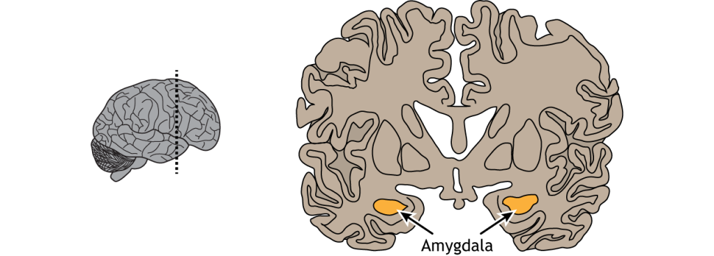
Figure 1. Drawing of the brain with the left and right amygdalae highlighted. Source: Amygdala’ by Casey Henley is licensed under a Creative Commons Attribution Non-Commercial Share-Alike (CC BY-NC-SA) 4.0 International License.
The authors analyzed how participant characteristics (such as age or sex) are associated with amygdala size. First, the size of the amygdala increased with age, which is indicative of typical development. Second, they found that the males’ amygdalae were larger on average, and that the amygdala was typically larger than the right. Lastly, males and females showed different trajectories of growth: in males, they saw that the amygdala rapidly grows earlier in development, followed by a slowed but steady continuation, whereas females’ amygdala demonstrated a slow and steady development across time.
They further conducted a series of standardized tests that assess infant and toddler development, including language, motor skills, communication, self-direction, social interaction, emotional expression, and self-regulation. For example, the infants and children were assessed on how they engage in social relationships, use emotions to interact in a meaningful way, use emotional gestures to communicate, and express emotions both positive (happy, excited, etc.) and negative (sad, angry, etc.). For more detailed information about the assessment used to assess development, please see this webpage.
Regarding the associations between amygdala size and brain function, several interesting patterns emerged: increased amygdala size was associated with decreased social and emotional development. Further, for males only, increased size of the left amygdala was associated with decreased receptive communication, or their ability to do such things as recognize sounds, make use of plurals or pronouns, understand instructions, and others. The authors suggest that these results corroborate previous research suggesting that infants with larger amygdalas tend to have impairments in social and communicative abilities.
In conclusion, the size of the amygdala may have a substantial impact on the development of understanding and interpreting social language or information. This study established a benchmark for understanding what typical amygdala development looks like, which in turn can be used to compare atypical developing amygdalae (plural for amygdala). For example, previous research has found that children with a diagnosis of Autism Spectrum Disorder typically have larger right amygdalae. Mueller et al. (2025) suggest that continued longitudinal research investigating amygdalae size and functional outcomes could mean that we can use the size of the amygdalae as a risk factor for worse developmental outcomes, which in turn would allow for earlier intervention/support. Future research is needed to understand the mechanisms underlying the association between amygdala size and social and emotional development; however, what is clear from these findings is that bigger brains aren’t always better brains!
Original Article: Mueller, M. E., Nichols, E. S., Al‐Saoud, S., De Vrijer, B., McKenzie, C. A., Eagleson, R., De Ribaupierre, S., & Duerden, E. G. (2025). Age‐Related Changes in the Amygdala From In Utero to Early Childhood: Association With Social and Cognitive Outcomes. Human Brain Mapping, 46(16), e70401. https://doi.org/10.1002/hbm.70401